Nearby my Australian town cemetery is a small watercourse. It’s a flowing mass of water molecules and colloids, propelled along the surface of the planet by gravitational force. This churning tends to move useful ions around the environment, making a nice heterogeneous soup of liquid, dissolved gasses and ions. More simply, a lot of water tends to result in a great habitat for life!
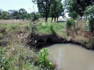
Given my interest in aquatic microorganisms, I collected a couple of water samples here for the purpose of observation. The location is — according to Wikipedia — a highland subtropical climate, so we’d expect to see common microorganisms that are ubiquitous to freshwater worldwide.
At the sampling site, freshwater algae was visible as green strands of biomass, later identified as masses of filamentous green algae. The samples were both collected from the southern bank via disposable pipettes and dispensed into sample containers and labelled. The first sample contained some of the algae that was visible to the naked eye, while the other contained some of the silt from the bank.
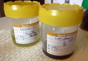
The sample on the left (sample 1) contains a small amount of the algal filaments, and was taken from the water directly next to the bank. The sample on the right (sample 2) contains evidence of silt and muddy water. I expect there to be lots of photosynthetic organisms in the sunlit waters of sample 1. Usually I can find lots of small protists swimming around the cover of larger organisms and plants.
Sample 1 contains what appears to be cyanobacteria — prokaryotic organisms that are capable of using solar radiation to produce energy. It is of interest to me that organisms like this produce most of the Earth’s atmospheric oxygen.
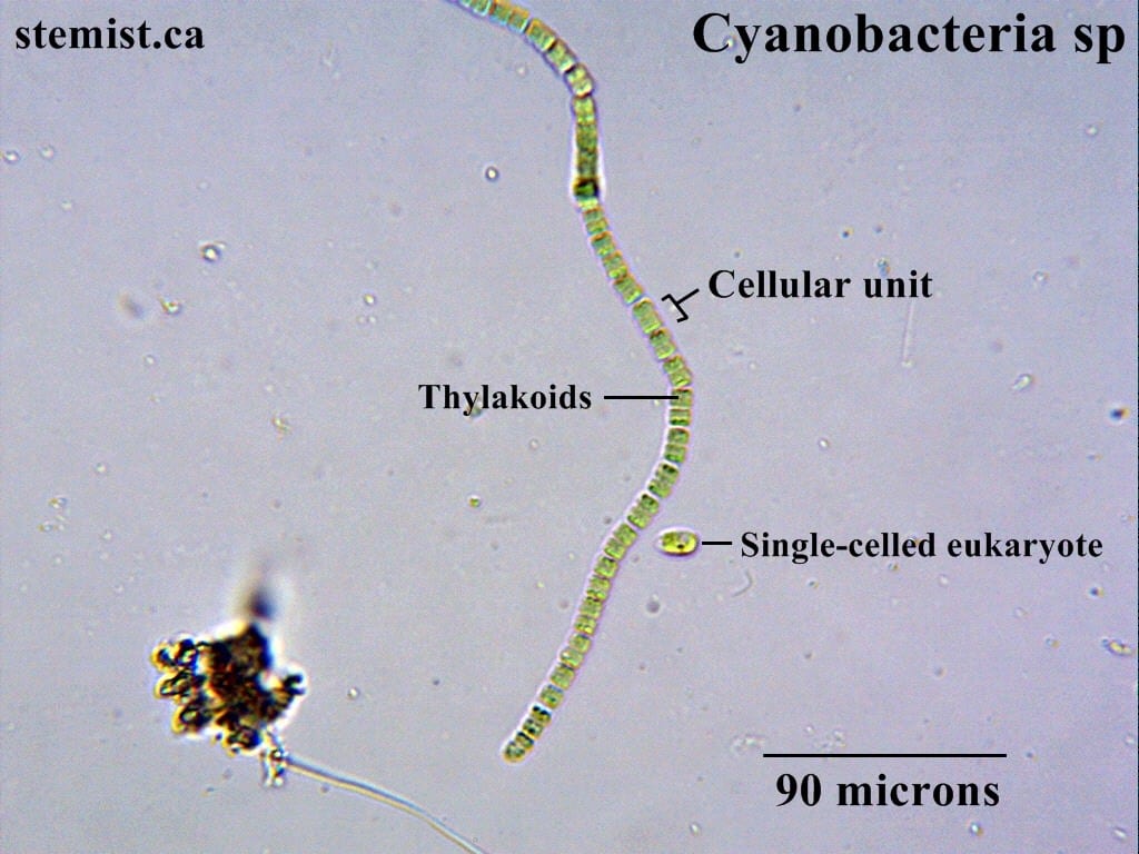
The cyanobacteria form long collections of individual cells. These filaments easily bundle up into enormous swaths of biomass. Within each cell are thylakoid compartments, which contain the biochemical machinery required to produce ATP. This process uses green-pigmented chlorophyll molecules, which absorb photons emitted from the sun to provide the energy required to do this. Now, all this is assuming that these cells are indeed a species of cyanobacteria. However, given their green and segmented appearance, it is probable that this is the case. There exists several different biochemical tests that could confirm this, such as API ID strips, if we really wanted to be sure.
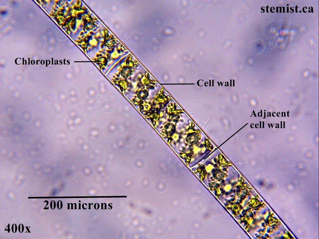
Very similar to the cyanobacteria are the large filamentous algae. They also form long chains of cells, but are much larger, and each cell has membrane-bound organelles. The filaments tend to bunch up in large collections of biomass that are visible to the naked eye.
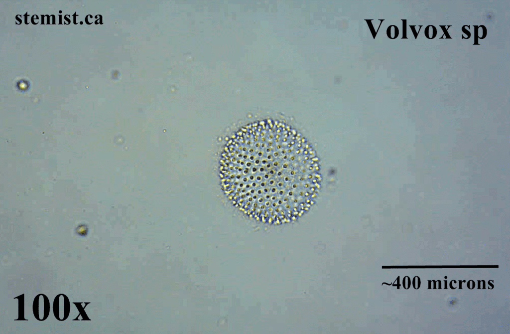
In addition to the above organisms, I also spotted a large number of volvox, which are microorganisms that form colonies of cells. Their flagella face outward, and the overall structure has some level of motility. In other words, it can move around. Below is an image I recorded several months ago that illustrates a volvox colony.
All these organisms exist on a scale that seems alien and different from our experience. However, we ourselves are familiar with reality on the scale of common measurements such as the metre, the kilogram, and others. It is interesting to think about how consciousness might understand the universe if we ourselves were on the scale of microscopic organisms.
This is part 1. Part 2 will contain more observations and pictures of organisms not described here. Stay tuned for more!